
Wilson's sign (osteochondritis dissecans of the medial femoral condyle)
Also called: Wilson test, Wilson's sign
For: Osteochondritis dissecans (medial femoral condyle)
A patch of subchondral bone weakens and may loosen or separate, with its overlying cartilage, from the medial femoral condyle at the lower end of the femur. It is seen mainly in older children, adolescents and young adults.
Not enough published figures to judgeShort answer: Not enough published figures to judge this test for osteochondritis dissecans (medial femoral condyle). The source cited below gives a sensitivity of 25% only.
How it is done
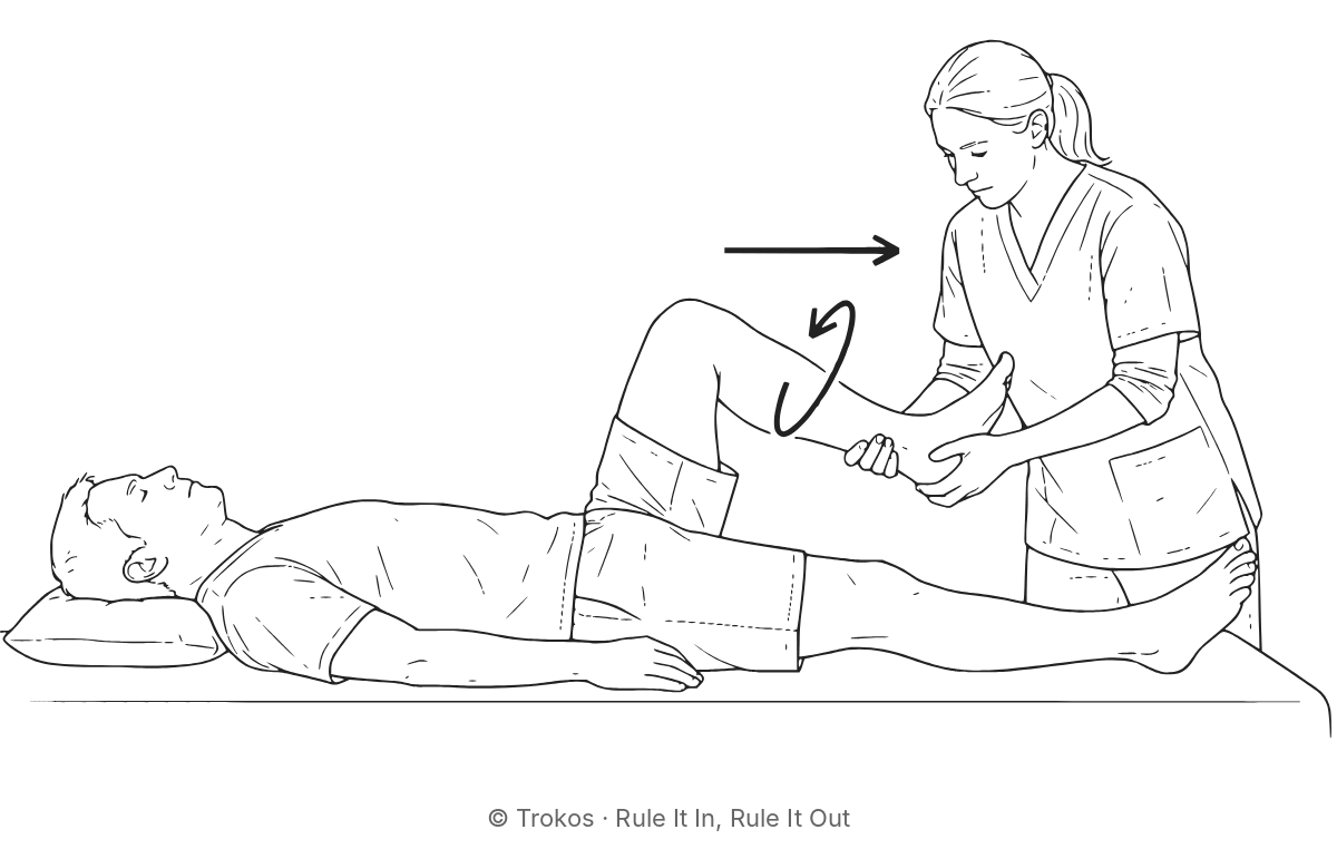
The patient lies supine with the knee at about 90°. The examiner internally rotates the tibia, then slowly extends the knee. Positive: Pain at around 30° of flexion that eases when the examiner turns the tibia outward.
What the numbers mean
- Too few published figures to say how much a result changes the picture.
Checked against the published abstract only; the full paper could not be accessed.
Study details: patients, setting, confidence intervals, what was read
| Study | Conrad 2003 (retrospective case series) |
|---|---|
| Patients | 32 patients; children and adolescents aged 9–17 with medial femoral osteochondritis dissecans on X-ray, from case records (no control group) |
| Compared against | radiographic osteochondritis dissecans |
| 95% confidence intervals | sensitivity 13–42% (calculated from the study’s counts) |
| What we read | Abstract only (full paper not available to us); PubMed abstract (PMID 12975201) and SAGE abstract page; full text restricted |
| Things to bear in mind | Small (32 patients), looked back through case records, and had no comparison group without the condition, so only sensitivity can be worked out. The abstract does not say who examined the patients or whether they knew the X-ray result. |
How these details are gathered and checked: Methods.
Source
Conrad JM, Stanitski CL. Osteochondritis dissecans: Wilson's sign revisited. Am J Sports Med. 2003;31(5):777–778. Journal article (DOI) · single study
Figures as they will appear in Rule It In, Rule It Out (Trokos, forthcoming). If a newer or better study changes them, this page changes with the book. Figures last updated from the book on 13 October 2026.
How to cite
Citing a figure? Cite the study it comes from. The main figures on this page come from:
Conrad JM, Stanitski CL. Osteochondritis dissecans: Wilson's sign revisited. Am J Sports Med. 2003;31(5):777–778.
Citing this page as a summary:
APA 7: Tompson, S. (2026, October 13). Wilson's sign (osteochondritis dissecans of the medial femoral condyle) for osteochondritis dissecans (medial femoral condyle). Trokos special tests library. Retrieved October 28, 2026, from https://trokos.com.au/tests/knee-wilsontest.html
Vancouver: Tompson S. Wilson's sign (osteochondritis dissecans of the medial femoral condyle) for osteochondritis dissecans (medial femoral condyle) [Internet]. Sunshine Coast (QLD): Trokos; 2026 [updated 2026 Oct 13; cited 2026 Oct 28]. Available from: https://trokos.com.au/tests/knee-wilsontest.html
Citing the whole library:
APA 7: Tompson, S. (2026). Trokos special tests library (Version November 2026) [Online reference library]. Zenodo. https://doi.org/10.5281/zenodo.23932041
Vancouver: Tompson S. Trokos special tests library [Internet]. Version November 2026. Geneva: Zenodo; 2026 [cited 2026 Nov 4]. Available from: https://doi.org/10.5281/zenodo.23932041
Get the book for the clinical reasoning behind each knee test it covers, and which ones to trust for the ACL, meniscus and kneecap
This page gives the published figures. Rule It In, Rule It Out adds captioned drawings with the exact positions, evidence dots, a fuller interpretation of each test, and the clinical reasoning on which tests to use and in what order.
Look inside the book: free sample pages
Coming November 2026: 330 pages, paperback and PDF. Launch offer: subscribers get the PDF for A$39 instead of A$49, for the first two weeks after publication. Be first to know at launch
One study, one test, once a month
Rule It In is a free evidence letter for musculoskeletal clinicians: one recent study explained, one examination test with its real numbers. From Trokos, publisher of Rule It In, Rule It Out.
Free when you confirm your email: a one-page PDF of the strongest special tests to rule in and rule out, with tests that carry a caution flagged separately.
Launch offer for subscribers: the PDF edition of Rule It In, Rule It Out for A$39 instead of A$49, for the first two weeks after publication. Sign up free to get the code on launch day.
By subscribing you agree to receive Rule It In by email, about once a month. You will get a confirmation email first, and nothing is sent until you click it. Your address is stored by Kit (Kit.com, United States) and used only for this newsletter and the subscriber launch offer; Kit records which issues are opened and which links are clicked. Unsubscribe with one click in any issue. Privacy.